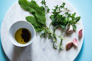
黃耆、當歸、三七水提物有效預防糖尿病視網膜病變
當歸補血湯,黃耆和當歸的水提物,是中醫治療缺血性疾病的常用方劑,在此修改添加三七(RRP),並在治療糖尿病方面進行了研究視網膜病變。所有這些草藥都具有抗炎特性,在中醫中經常用於治療糖尿病和糖尿病併發症 [ 1 , 2 ]。我們在這裡表明,RRP 治療可防止白細胞粘附在血管壁上,減弱血管滲漏,抑制脫細胞毛細血管的形成,這是糖尿病視網膜病變的三種早期標誌性病理,從而改善了糖尿病大鼠模型中的視網膜損傷並阻止了 DR 進展。RRP 對糖尿病視網膜病變的保護可能是通過抑制炎症反應和改善視網膜微循環來實現的。
DR 是一種低度慢性炎症性疾病 [ 3 ]。以前的研究表明,抗炎藥物可以預防早期 DR [ 4 , 5 ]。黃芪、當歸和三七已被證明可抑制促炎因子或趨化因子的表達,如 IL- 1β、IL-6、TNF- α、VEGF、MCP-1、ICAM-1 或 VCAM-1 [ 6,7 – 9 , 10 , 11]。在這裡,我們證實 RRP 抑制了一系列促炎細胞因子的基因表達,這可能因此導致糖尿病大鼠視網膜炎症減少。促炎細胞因子(如 TNF- α和白細胞介素)和趨化因子(如 MCP-1)激活內皮以增加粘附分子(如 ICAM-1、VCAM-1)和趨化因子的表達,從而介導白細胞/單核細胞附著在血管壁上並通過內皮遷移 [ 12 ]。ICAM-1 阻斷劑可防止糖尿病視網膜白細胞淤滯和血管滲漏 [ 15]。我們推測 RRP 可以通過阻斷炎症和隨後激活白細胞/單核細胞粘附過程來保護視網膜免受白細胞淤滯。經過12週的給藥,我們觀察到RRP抑制炎性基因的表達和NF- κ的活化和ICAM-1和VCAM-1的水平的efficaciously逆轉糖尿病誘導的高程。白細胞淤滯增加是 DR的早期事件,被認為會導致 BRB 分解、內皮細胞損傷和毛細血管丟失 [ 16 ]。在本研究中,我們觀察到糖尿病 GK 大鼠視網膜血管內皮上粘附的白細胞/單核細胞數量增加;因此,糖尿病 GK 大鼠的血管通透性和脫細胞毛細血管數量顯著增加。RRP治療抑制了視網膜血管系統中的白細胞淤滯,這可能受益於ICAM-1和VCAM-1分子的抑製表達。內部 BRB 的改變是 DR 的標誌,在 GK 大鼠 [ 17 ] 以及糖尿病患者 [ 18 , 19 ]的視網膜病變進展早期,BRB 的分解出現]。與減弱的白細胞淤滯相一致,我們發現,與未治療的糖尿病組相比,12週的RRP應用顯著降低了糖尿病GK大鼠的視網膜血管通透性並抑制了脫細胞毛細血管的形成。此外,RRP 的 3 週應用大大減少了 STZ 誘導的糖尿病大鼠的視網膜血管滲漏,表明 RRP 在預防糖尿病視網膜損傷方面具有整體保護作用。我們沒有看到 RRP 對體重、血糖和 TG 或 TC 水平的明顯影響而視網膜病變的改善是明顯的,這意味著高血糖引起的局部環境變化尤其是炎症在視網膜病變的發病機制中比全身性高血糖起著更重要的作用。已經表明,視網膜炎症介質是高血糖停止後視網膜病變抵抗停止的主要因素,並且在血糖控制良好至少六個月後,促炎細胞因子和粘附分子仍保持高水平,隨後六個月血糖控制不佳[ 20 ] . 高血糖的逆轉不能對作為 DR 特徵的組織病理學的發展產生任何顯著影響 [ 21 ]。各種研究表明,抑制促炎細胞因子(如 IL-1β、TNF- α和 VEGF)、趨化因子(如 MCP-1)或粘附分子(如 ICAM-1)可以減少糖尿病引起的白細胞淤滯 [ 3 , 15 ]、視網膜毛細血管的退化 [ 22 ] ,或 BRB 故障 [ 23 ]。因此,我們的結果表明,RRP 的抗視網膜病變作用可能與降低血糖無關,而是由於抑制了 GK 大鼠視網膜中的炎症。除了在糖尿病 GK 大鼠的體內研究外,我們還證實了炎性細胞因子對培養的內皮細胞的 RRP 抑製作用。
內皮細胞凋亡導致的內皮細胞丟失是DR的早期病理變化;我們在此表明,在培養的 EA.hy926 細胞中,RRP 逆轉了受抑制的細胞遷移並減少了由高糖誘導的細胞死亡,表明 RRP 具有預防早期內皮細胞死亡的保護作用。
總之,我們的結果表明 RRP 對 GK 大鼠 DR 的發展和/或進展具有抑製作用。RRP 預防視網膜病變可能與抑制白細胞淤滯和炎症有關。糖尿病患者通常只有在視力並發症已經發生後才去看眼科醫生,此時沒有太多理想的干預措施可以逆轉或消除損害。RRP 在改善 DR 增加的通透性、白細胞淤滯和脫細胞毛細血管等特徵方面具有很強的作用,可作為 DR 管理的預防和/或乾預策略。
[ 1 ] H. Zhang, S. Chen, X. Deng, X. Yang, and X. Huang, “Danggui-Buxue-Tang decoction has an anti-inflammatory effect in diabetic atherosclerosis rat model,” Diabetes Research and Clinical Practice, vol. 74, no. 2, pp. 194–196, 2006.
[ 2 ] C. Y. Yang, J. Wang, Y. Zhao et al., “Anti-diabetic effects of Panax notoginseng saponins and its major anti-hyperglycemic components,” Journal of Ethnopharmacology, vol. 130, no. 2, pp. 231–236, 2010.
[ 3 ] S. Rangasamy, P. G. McGuire, and A. Das, “Diabetic retinopathy and inflammation: novel therapeutic targets,” Middle East African Journal of Ophthalmology, vol. 19, no. 1, pp. 52–59, 2012.
[ 4 ]A. M. Joussen, V. Poulaki, N. Mitsiades et al., “Nonsteroidal anti-inflammatory drugs prevent early diabetic retinopathy via TNF-alpha suppression,” The FASEB Journal, vol. 16, no. 3, pp. 438–440, 2002.
[ 5 ] P. Pakneshan, A. E. Birsner, I. Adini, C. M. Becker, and R. J. D’Amato, “Differential suppression of vascular permeability and corneal angiogenesis by nonsteroidal anti-inflammatory drugs,” Investigative Ophthalmology and Visual Science, vol. 49, no. 9, pp. 3909–3913, 2008.
[ 6 ] M. Ryu, E. H. Kim, M. Chun et al., “Astragali Radix elicits anti-inflammation via activation of MKP-1, concomitant with attenuation of p38 and Erk,” Journal of Ethnopharmacology, vol. 115, no. 2, pp. 184–193, 2008.
[ 7 ] C. Yang, S. Niu, L. Yu, S. Zhu, J. Zhu, and Q. Zhu, “The aqueous extract of Angelica sinensis, a popular Chinese herb, inhibits wear debris-induced inflammatory osteolysis in mice,” Journal of Surgical Research, vol. 176, no. 2, pp. 476–483, 2012.
[ 8 ] C. Han and J. Guo, “Antibacterial and anti-inflammatory activity of traditional Chinese herb pairs, Angelica sinensis and Sophora flavescens,” Inflammation, vol. 35, no. 3, pp. 913–919, 2012.
[ 9 ] W. W. Chao, Y. H. Hong, M. L. Chen, and B. F. Lin, “Inhibitory effects of Angelica sinensis ethyl acetate extract and major compounds on NF-κB trans-activation activity and LPS-induced inflammation,” Journal of Ethnopharmacology, vol. 129, no. 2, pp. 244–249, 2010.
[ 10 ] A. Rhule, S. Navarro, J. R. Smith, and D. M. Shepherd, “Panax notoginseng attenuates LPS-induced pro-inflammatory mediators in RAW264.7 cells,” Journal of Ethnopharmacology, vol. 106, no. 1, pp. 121–128, 2006.
[ 11 ] L. Dou, Y. Lu, T. Shen et al., “Panax notogingseng saponins suppress RAGE/MAPK signaling and NF-kappaB activation in apolipoprotein-E-deficient atherosclerosis-prone mice,” Cellular Physiology and Biochemistry, vol. 29, no. 5-6, pp. 875–882, 2012.
[ 12 ] G. I. Liou, “Diabetic retinopathy: role of inflammation and potential therapies for anti-inflammation,” World Journal of Diabetes, vol. 1, no. 1, pp. 12–18, 2010.
[ 13 ] J. A. Vincent and S. Mohr, “Inhibition of caspase-1/interleukin-1β signaling prevents degeneration of retinal capillaries in diabetes and galactosemia,” Diabetes, vol. 56, no. 1, pp. 224–230, 2007.
[ 14 ] A. M. Joussen, S. Doehmen, M. L. Le et al., “TNF-α mediated apoptosis plays an important role in the development of early diabetic retinopathy and long-term histopathological alterations,” Molecular Vision, vol. 15, pp. 1418–1428, 2009.
[ 16 ] M. V. Van Hecke, J. M. Dekker, G. Nijpels et al., “Inflammation and endothelial dysfunction are associated with retinopathy: the Hoorn Study,” Diabetologia, vol. 48, no. 7, pp. 1300–1306, 2005.
[ 17 ] A. Carmo, J. G. Cunha-Vaz, A. P. Carvalho, and M. C. Lopes, “Nitric oxide synthase activity in retinas from non-insulin-dependent diabetic Goto-Kakizaki rats: correlation with blood-retinal barrier permeability,” Nitric Oxide, vol. 4, no. 6, pp. 590–596, 2000.
[ 18 ] S. E. Moss, R. Klein, and B. E. K. Klein, “The 14-year incidence of visual loss in a diabetic population,” Ophthalmology, vol. 105, no. 6, pp. 998–1003, 1998.
[ 19 ] A. Joussen, N. Smyth, and C. Niessen, “Pathophysiology of diabetic macular edema,” Developments in Ophthalmology, vol. 39, pp. 1–12, 2007.
[ 20 ] P. S. Chan, M. Kanwar, and R. A. Kowluru, “Resistance of retinal inflammatory mediators to suppress after reinstitution of good glycemic control: novel mechanism for metabolic memory,” Journal of Diabetes and its Complications, vol. 24, no. 1, pp. 55–63, 2010.
[ 21 ] R. A. Kowluru, M. Kanwar, and A. Kennedy, “Metabolic memory phenomenon and accumulation of peroxynitrite in retinal capillaries,” Experimental Diabesity Research, vol. 2007, Article ID 21976, 7 pages, 2007.
[ 22 ] J. A. Vincent and S. Mohr, “Inhibition of caspase-1/interleukin-1β signaling prevents degeneration of retinal capillaries in diabetes and galactosemia,” Diabetes, vol. 56, no. 1, pp. 224–230, 2007.
[ 23 ] A. M. Joussen, S. Doehmen, M. L. Le et al., “TNF-α mediated apoptosis plays an important role in the development of early diabetic retinopathy and long-term histopathological alterations,” Molecular Vision, vol. 15, pp. 1418–1428, 2009.
中醫藥治療老年性黃斑變性
S Wang, K Cunnusamy – Expert opinion on therapeutic patents, 2013
當歸可以減少氧化應激和炎症。具體而言,當歸顯示在內毒素誘導的葡萄膜炎中減少 NF-κB 活化和炎症基因表達 ( 1 )。當歸的多醣成分可以通過抑製過量氮氧化物 (NO) 和活性氧 (ROS) 的釋放來防止巨噬細胞介導的損傷 (2 )。當歸的提取物可以是促血管生成或抗血管生成的,具體取決於配方 ( 3 , 4 )。
在膠原誘導的關節炎模型中,三七提取物顯示出抑制炎症基因表達和炎症的作用 ( 5 , 6 )。它還通過抑制黃嘌呤氧化酶活性發揮抗氧化作用 ( 7 )。
Poria cocos wolf在接觸性皮炎的臨床研究中顯示出抗炎特性 ( 8 )。來自茯苓菌絲體的多醣也顯示出抗腫瘤作用 ( 9 )。澱粉樣蛋白 β 與 AMD 的發病機制有關 ( 10 )。在這種情況下,茯苓狼也可能對乾性 AMD 有益,因為茯苓和當歸 Nakai 的組合已被證明可以防止小鼠 β-澱粉樣蛋白斑塊沉積和阿爾茨海默病樣病理 ( 11 )。
- Han C, Guo J. Antibacterial and anti-inflammatory activity of traditional Chinese herb pairs, Angelica sinensis and Sophora flavescens. Inflammation. 2012;35(3):913–919.
- Yang X, et al. Component and antioxidant properties of polysaccharide fractions isolated from Angelica sinensis (OLIV.) DIELS. Biological & pharmaceutical bulletin. 2007;30(10):1884–1890.
- Lam HW, et al. The angiogenic effects of Angelica sinensis extract on HUVEC in vitro and zebrafish in vivo. Journal of cellular biochemistry. 2008;103(1):195–211.
- Yeh JC, et al. The natural compound n-butylidenephthalide derived from the volatile oil of Radix Angelica sinensis inhibits angiogenesis in vitro and in vivo. Angiogenesis. 2011;14(2):187–197.
- Chang SH, et al. Anti-inflammatory effects of BT-201, an n-butanol extract of Panax notoginseng, observed in vitro and in a collagen-induced arthritis model. Clin Nutr. 2007;26(6):785–791.
- Jang YJ, Kim ME, Ko SY. n-Butanol extracts of Panax notoginseng suppress LPS-induced MMP-2 expression in periodontal ligament fibroblasts and inhibit osteoclastogenesis by suppressing MAPK in LPS-activated RAW264.7 cells. Archives of oral biology. 2011;56(11):1319–1327.
- Choi RC, et al. Anti-oxidative effects of the biennial flower of Panax notoginseng against H2O2-induced cytotoxicity in cultured PC12 cells. Chinese medicine. 2010;5:38.
- Fuchs SM, et al. Assessment of anti-inflammatory activity of Poria cocos in sodium lauryl sulphate-induced irritant contact dermatitis. Skin Res Technol. 2006;12(4):223–227.
- Kanayama H, Adachi N, Togami M. A new antitumor polysaccharide from the mycelia of Poria cocos wolf. Chemical & pharmaceutical bulletin. 1983;31(3):1115–1118.
- Ding JD, et al. Anti-amyloid therapy protects against retinal pigmented epithelium damage and vision loss in a model of age-related macular degeneration. Proceedings of the National Academy of Sciences of the United States of America. 2011;108(28):E279–287.
- Seo JS, et al. A modified preparation (LMK03) of the oriental medicine Jangwonhwan reduces Abeta(1-42) level in the brain of Tg-APPswe/PS1dE9 mouse model of Alzheimer disease. Journal of ethnopharmacology. 2010;130(3):578–585.